
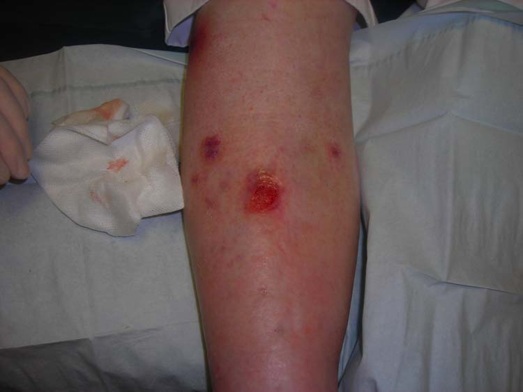
Image: "Venous ulcer dorsal leg" by Jonathan Moore from the case series "Creating the Ideal Microcosm for Rapid Incorporation of Bioengineered Alternative Tissues Using An Advanced Hydrogel Impregnated Gauze Dressing: A Case Series", The Foot and Ankle Online Journal 1 (9): 2, is licensed under CC BY-SA 3.0. Link to the source.
Leg Ulcers
Introduction | Aetiology and Risk Factors | Clinical Presentation | Diagnosis | Management and Treatment | Prevention | When to Refer | References
Introduction
Leg ulcers are chronic wounds that occur on the lower limbs due to various underlying conditions, most commonly venous insufficiency, arterial disease, or a combination of both. These ulcers can cause significant morbidity and are often difficult to heal without appropriate management. They are more prevalent in older adults and can have a substantial impact on quality of life due to pain, infection risk, and impaired mobility.
Aetiology and Risk Factors
Leg ulcers are classified based on their underlying cause:
- Venous Ulcers: The most common type, accounting for about 70% of leg ulcers. They are typically caused by chronic venous insufficiency, which leads to increased pressure in the veins, resulting in skin breakdown and ulceration.
- Arterial Ulcers: These occur due to poor arterial blood flow, often as a result of peripheral arterial disease (PAD). They are less common but more challenging to treat due to compromised blood supply.
- Mixed Venous-Arterial Ulcers: These ulcers have both venous and arterial components, complicating treatment and requiring careful management to balance wound healing and blood flow.
- Other Causes: Less common causes of leg ulcers include diabetes (neuropathic ulcers), vasculitis, malignancy, and trauma.
Risk factors for leg ulcers include:
- Age: Older adults are at higher risk due to decreased mobility, venous insufficiency, and arterial disease.
- Obesity: Increased weight puts additional pressure on the veins, exacerbating venous insufficiency.
- Smoking: Smoking impairs blood flow and increases the risk of arterial disease.
- History of Deep Vein Thrombosis (DVT): Previous DVT can damage venous valves, leading to chronic venous insufficiency.
- Diabetes: Poorly controlled diabetes can lead to neuropathy and peripheral arterial disease, increasing the risk of ulceration.
- Immobility: Reduced mobility can lead to venous stasis and increased risk of ulceration.
Clinical Presentation
Leg ulcers present with various clinical features depending on their underlying cause:
1. Venous Ulcers
- Location: Typically located on the medial malleolus (inside of the ankle).
- Appearance: Shallow, irregularly shaped ulcers with a granulating base, often surrounded by brownish skin discolouration (haemosiderin staining) and lipodermatosclerosis.
- Exudate: Venous ulcers tend to be moist with moderate to heavy exudate.
- Symptoms: Patients may report aching, heaviness, and swelling of the legs, which worsens with prolonged standing and improves with elevation.
2. Arterial Ulcers
- Location: Commonly found on the toes, feet, or lateral malleolus (outside of the ankle).
- Appearance: Well-demarcated, "punched-out" lesions with a pale or necrotic base. The surrounding skin may be cool, shiny, and hairless.
- Exudate: Typically dry with minimal exudate.
- Symptoms: Patients often experience severe pain, particularly at night or when the leg is elevated (rest pain). They may also have signs of peripheral arterial disease, such as diminished pulses.
3. Mixed Venous-Arterial Ulcers
- Clinical Features: These ulcers have characteristics of both venous and arterial ulcers, making them more complex to manage. They may present with mixed symptoms and appearances.
4. Diabetic/Neuropathic Ulcers
- Location: Often found on pressure points such as the heels, metatarsal heads, or tips of the toes.
- Appearance: Painless, deep ulcers with a punched-out appearance. The surrounding skin may be calloused.
- Symptoms: Due to neuropathy, patients may not feel pain even with deep ulcers, which can delay diagnosis and treatment.
Diagnosis
The diagnosis of leg ulcers involves a combination of clinical assessment and diagnostic investigations:
- History: Take a detailed history, including the onset and duration of the ulcer, previous episodes, risk factors, and any associated symptoms (e.g., claudication, rest pain, swelling).
- Physical Examination: Inspect the ulcer and surrounding skin, palpate peripheral pulses, and assess for signs of venous insufficiency (e.g., varicose veins, haemosiderin staining) or arterial disease (e.g., cool, shiny skin).
- Ankle-Brachial Pressure Index (ABPI): Measure the ABPI to assess arterial blood flow. An ABPI of <0.9 suggests arterial insufficiency, while an ABPI >1.3 may indicate calcified, non-compressible vessels, often seen in diabetes.
- Doppler Ultrasound: Duplex ultrasound can help evaluate venous insufficiency and arterial blood flow.
- Wound Swab and Culture: If there is clinical evidence of infection (e.g., increased exudate, erythema, odour), swab the ulcer for culture and sensitivity.
- Blood Tests: Perform routine blood tests, including a full blood count (FBC), glucose levels, and HbA1c for diabetic patients, to assess for underlying conditions that may impede healing.
Management and Treatment
The management of leg ulcers depends on the underlying cause and involves wound care, addressing the cause, and promoting healing:
1. Wound Care
- Cleansing: Clean the ulcer with saline or a mild antiseptic solution. Avoid harsh chemicals that can delay healing.
- Debridement: Remove necrotic tissue to promote healing. This can be done surgically, mechanically, enzymatically, or autolytically depending on the ulcer and patient condition.
- Dressings: Choose dressings based on the ulcer type and exudate level. For venous ulcers, use moisture-retentive dressings such as hydrogels or foam dressings. Arterial ulcers may benefit from dry, non-adhesive dressings.
- Compression Therapy: Compression bandaging or stockings are essential for venous ulcers to reduce venous hypertension. Ensure arterial disease is excluded before applying compression.
- Infection Control: Use topical antimicrobials or systemic antibiotics if there is clinical evidence of infection.
2. Managing the Underlying Cause
- Venous Ulcers: Compression therapy is the mainstay of treatment, along with lifestyle modifications such as weight loss, leg elevation, and increased physical activity. Consider referral for venous surgery if indicated.
- Arterial Ulcers: Optimise arterial blood flow with antiplatelet agents, statins, and lifestyle changes (e.g., smoking cessation). Severe cases may require revascularisation procedures.
- Diabetic Ulcers: Strict glycaemic control, regular foot care, and offloading pressure from the ulcer site (e.g., through specialised footwear or orthotics) are crucial.
3. Pain Management
- Analgesics: Provide appropriate pain relief with paracetamol, NSAIDs, or opioids for severe pain. Consider topical analgesics for neuropathic pain in diabetic ulcers.
4. Nutrition and Support
- Nutrition: Ensure adequate nutritional support, especially in elderly or malnourished patients, to promote wound healing. Supplement with vitamins and minerals as necessary.
- Psychosocial Support: Address the psychological impact of chronic ulcers, which can affect quality of life. Consider referral to a support group or counsellor if needed.
Prevention
Preventing leg ulcers and their recurrence involves managing risk factors and ongoing care:
- Compression Therapy: Long-term use of compression stockings is crucial for preventing recurrence of venous ulcers.
- Lifestyle Modifications: Encourage weight management, smoking cessation, regular exercise, and leg elevation to reduce venous hypertension.
- Foot Care: For diabetic patients, regular foot checks, appropriate footwear, and good glycaemic control are essential to prevent ulceration.
- Regular Monitoring: Schedule regular follow-ups to monitor ulcer healing and adjust treatment as needed.
When to Refer
Referral to a specialist, such as a dermatologist, vascular surgeon, or diabetic foot clinic, may be necessary in the following situations:
- Non-Healing Ulcers: If an ulcer fails to improve with standard care after 4-6 weeks.
- Suspected Arterial Ulcers: For further evaluation and potential revascularisation procedures.
- Infection: Severe or spreading infection that may require systemic antibiotics or surgical intervention.
- Uncertain Diagnosis: If the cause of the ulcer is unclear or if there is suspicion of an underlying malignancy.
- Complex Cases: Mixed venous-arterial ulcers, diabetic ulcers with complications, or ulcers with significant psychosocial impact.
References
- National Institute for Health and Care Excellence (2024) Chronic Wounds: Leg Ulcers. Available at: https://www.nice.org.uk/guidance/ng147 (Accessed: 26 August 2024).
- British Association of Dermatologists (2024) Guidelines for the Management of Leg Ulcers. Available at: https://www.bad.org.uk (Accessed: 26 August 2024).
- British National Formulary (2024) Wound Management and Dressings. Available at: https://bnf.nice.org.uk/ (Accessed: 26 August 2024).

Check out our YouTube channel
Blueprint Page
Explore the comprehensive blueprint for Physician Associates, covering all essential topics and resources.
Book Your Session
Enhance your skills with personalised tutoring sessions tailored for Physician Associates.