Deep Vein Thrombosis (DVT)
Definition | Aetiology | Pathophysiology | Risk Factors | Signs and Symptoms | Investigations | Management | Patient Advice
Definition
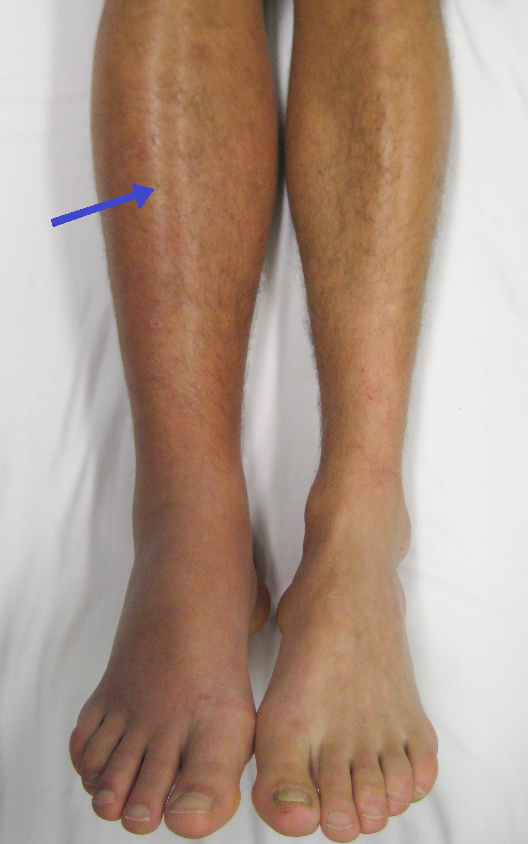
Deep Vein Thrombosis (DVT) is the formation of a blood clot in a deep vein, most commonly in the legs. It can cause pain and swelling and carries a risk of life-threatening complications such as pulmonary embolism (PE). See Figure 1.
Aetiology
DVT results from a combination of factors that contribute to clot formation, known as Virchow's triad:
- Venous Stasis: Reduced blood flow in veins due to immobility, prolonged bed rest, or long-haul flights.
- Endothelial Injury: Damage to the vein wall from trauma, surgery, or inflammation.
- Hypercoagulability: Increased clotting tendency due to conditions such as pregnancy, cancer, or inherited thrombophilias (e.g., Factor V Leiden).
Pathophysiology
DVT develops due to the following processes:
- Clot Formation: Platelets and fibrin aggregate to form a thrombus within the vein.
- Venous Obstruction: The clot obstructs blood flow, leading to swelling and increased venous pressure.
- Risk of Embolisation: The clot may dislodge and travel to the lungs, causing pulmonary embolism.
Risk Factors
Key risk factors include:
- Prolonged immobility (e.g., after surgery or long travel).
- Recent surgery, particularly orthopaedic or pelvic procedures.
- Pregnancy or postpartum period.
- Cancer or chemotherapy.
- Use of oestrogen-containing contraceptives or hormone replacement therapy.
- Obesity or smoking.
- Inherited thrombophilias (e.g., Factor V Leiden mutation).
Signs and Symptoms
Symptoms of DVT include:
- Swelling: Unilateral swelling of the affected leg.
- Pain: Calf or thigh pain, often described as cramping or aching.
- Redness and Warmth: Over the affected area.
- Homan's Sign: Pain in the calf on dorsiflexion of the foot (not specific).
Investigations
Key investigations and common positive findings include:
- Wells Score: A clinical scoring system to assess DVT risk.
- D-dimer Test: Elevated levels indicate active clot formation and breakdown, though not specific for DVT.
- Compression Ultrasound: The diagnostic test of choice. Positive findings include non-compressibility of the affected vein.
- Venography: Used in complex cases to visualise the clot directly, though rarely needed.
Management
1. Primary Care Management
- Immediate Referral: Urgent referral to secondary care if DVT is suspected.
- Initial Anticoagulation: If available, administer low-molecular-weight heparin (e.g., enoxaparin) while awaiting secondary care review.
2. Secondary Care Management
- Anticoagulation Therapy: Start with low-molecular-weight heparin, transitioning to oral anticoagulants such as apixaban or rivaroxaban.
- Monitoring: Regular INR monitoring if using warfarin.
- Thrombolysis: Considered in massive DVT or phlegmasia cerulea dolens, typically performed by an interventional radiologist.
- Inferior Vena Cava (IVC) Filter: For patients with contraindications to anticoagulation, performed by a vascular specialist.
Patient Advice
Key advice includes:
- Take anticoagulants as prescribed and attend regular follow-ups.
- Stay mobile and avoid prolonged immobility.
- Maintain a healthy weight and stop smoking.
- Wear compression stockings to prevent post-thrombotic syndrome, if recommended.
- Seek immediate medical attention for symptoms of pulmonary embolism, such as sudden chest pain or breathlessness.
Figure 1

Image showing a deep vein thrombosis in the right leg, with swelling and redness.
References
- James Heilman, MD (2015). Deep Vein Thrombosis of the Right Leg [Image]. Available at: https://upload.wikimedia.org/wikipedia/commons/2/21/Deep_vein_thrombosis_of_the_right_leg.jpg (Accessed: 30 December 2024).
| Clinical Feature | Points |
|---|---|
| Active cancer | 1 |
| Paralysis, paresis, or immobilization of lower extremity | 1 |
| Recently bedridden for more than 3 days, or major surgery within the past 12 weeks | 1 |
| Localized tenderness along the distribution of the deep venous system | 1 |
| Entire leg swelling | 1 |
| Calf swelling at least 3 cm larger than asymptomatic side (measured 10 cm below tibial tuberosity) | 1 |
| Pitting edema confined to symptomatic leg | 1 |
| Collateral non-varicose veins | 1 |
| Previously diagnosed DVT or PE | 1 |
Interpretation:
A score of 0 or less: low probability of DVT
A score of 1-2: moderate probability of DVT
A score of 3 or more: high probability of DVT
If suspected DVT refer to local DVT clinic
